

Electron Microscopy Laboratory
Head of the laboratory: Prof. Dr hab. Marek Awdankiewicz,
e-mail: marek.awdankiewicz/at/uwr.edu.pl
Electron Microscopy Laboratory
Institute of Geological Sciences of the University of Wrocław
ul. Cybulskiego 30, 50-205 Wrocław
tel. 71 375 92 91
Description of activities
The apparatus available at EML allows for the examination of rocks, minerals, and other solid materials (including metals, alloys, mineral resources, gemstones, building materials, solid waste, slag, atmospheric dust, and other environmental samples) and fluid inclusions in solid materials.
The Jeol JSM-IT500LA scanning electron microscope with equipment enables:
- operation in low and high vacuum conditions,
- Observations over a magnification range of 5x to 300,000x, with a resolution from 3 nm at 30 kV to 15 nm at 1kV,
- imaging of studied substances using secondary electron (SE) techniques, back-scattered electrons (BSE), and cathodoluminescence (CL),
- qualitative and semi-quantitative chemical composition analyses using the energy dispersion (EDS) techniques, including point analysis, profiles and chemical composition maps.
The Raman spectrometer enables the execution of structural-chemical analyzes of various substances, their identification, making maps of spatial differentiation, and others using a confocal microscope.
Equipment of the Laboratory
Scanning electron microscopeJeol JSM-IT500LA

| Equipped with: |
| – Secondary Electrons (SE) Detector, |
| – Back-Scattered Electrons (BSE) Detector, |
| – Cathodoluminescence (CL ) Detector, |
| – Energy-Dispersive Spectrometer (EDS), |
| – Structural-Chemical Analyzer (SCA) Interface for Raman Spectroscopy. |
Raman Spectrometer
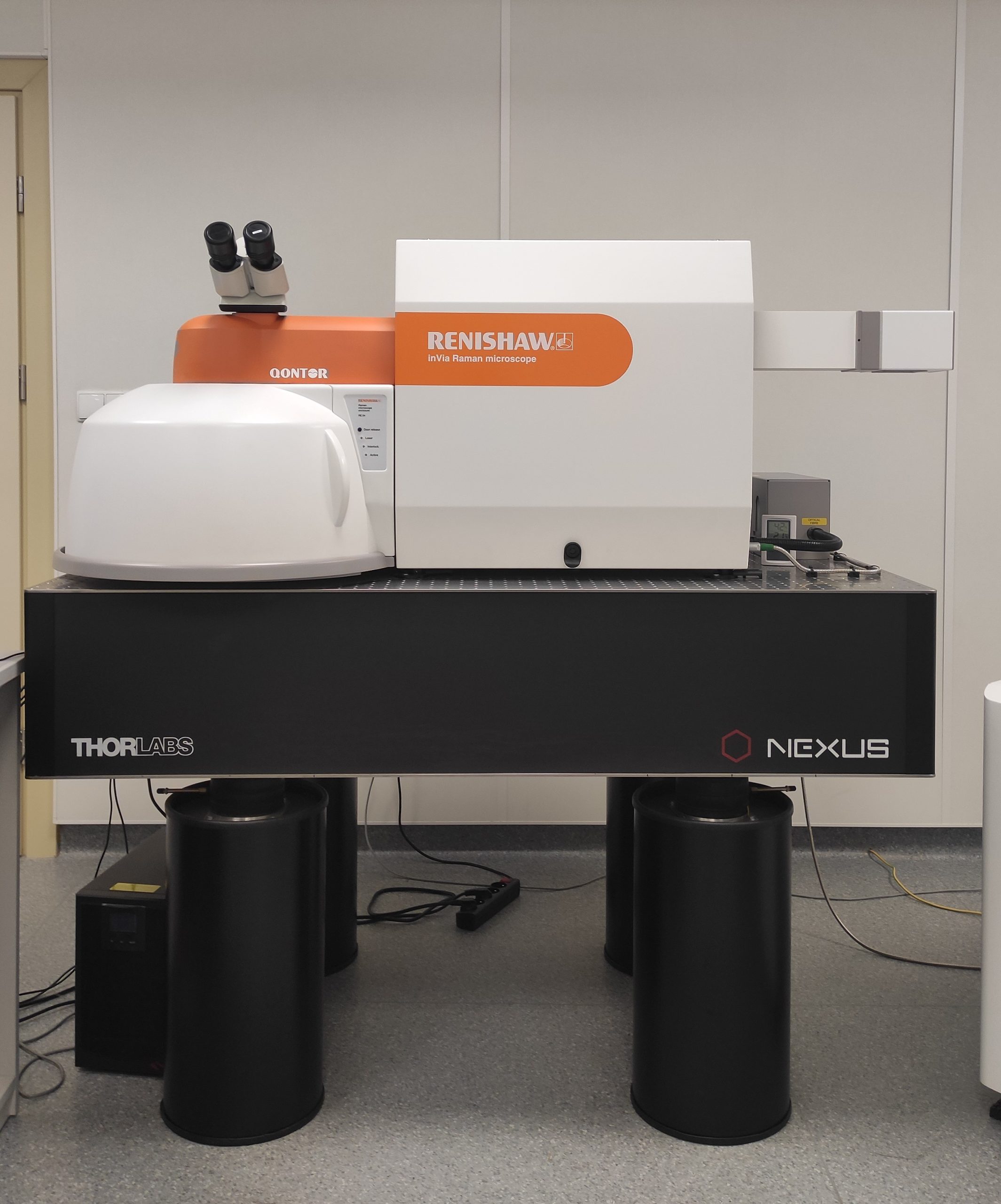
| Coupled to a scanning microscope and a confocal optical microscope, Renishaw inVia Qontor model, equipped with 785 nm and 532 nm diode lasers. |
| Combining a Raman spectroscope with a scanning microscope via an SCM interface allows for comprehensive, co-located characterization of the physical and chemical characteristics of the samples under study directly in the SEM chamber, including substance identification, imaging of morphology, textural features, chemical differentiation, and elemental composition. |
Price list for services using the scanning electron microscope and/or Raman spectrometer:
– research in cooperation with the staff of the Institute of Geological Sciences of the University of Wrocław PLN 140/hour + VAT
– commercial/other orders PLN 280/hour + VAT

